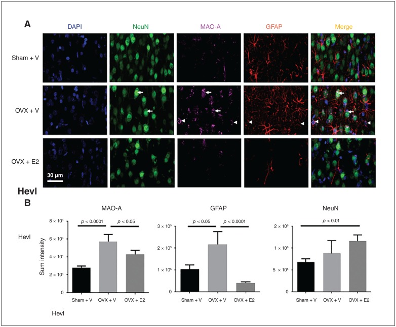
Fig. 5.
E2 reduces OVX-induced oxidative stress and glial cell activation in rats (6 d post-OVX). (A) Representative immunofluorescence images of dorsal raphe of OVX female rats treated with vehicle, OVX + vehicle and OVX + E2. Nuclei were counterstained with DAPI (blue). Note the nuclear staining of NeuN (green), cytosol punctate staining of MAO-A (magenta) and glial cell–specific staining of GFAP (red). Also note the even distribution of the NeuN staining in the 3 conditions, while both MAO-A and GFAP expressions were increased in OVX rats. Scale bar = 50 μm. Arrows and arrowheads indicate the representative cells expressing MAO-A + NeuN and MAO-A + GFAP, respectively. (B) The immunoreactive intensities of MAO-A, GFAP and NeuN in the dorsal raphe of sham + vehicle, OVX + vehicle and OVX + E2 female rats, analyzed using 1-way ANOVA and a subsequent Bonferroni post hoc test. Note the significant increases of MAO-A (F2,3110 = 8.80, p < 0.001) and GFAP (F2,8082 = 11.71, p < 0.001) expression in the dorsal raphe of OVX rats, but OVX did not change the expression of NeuN (F2,2655 = 1.73, p = 0.18); E2 significantly reduced OVX-induced MAO-A and GFAP expression. Although OVX did not change NeuN expression, E2 increased NeuN expression in OVX rats v. sham + vehicle rats. ANOVA = analysis of variance; E2 = estradiol; MAO-A = monoamine oxidase A; OVX = ovariectomy; V = vehicle.

