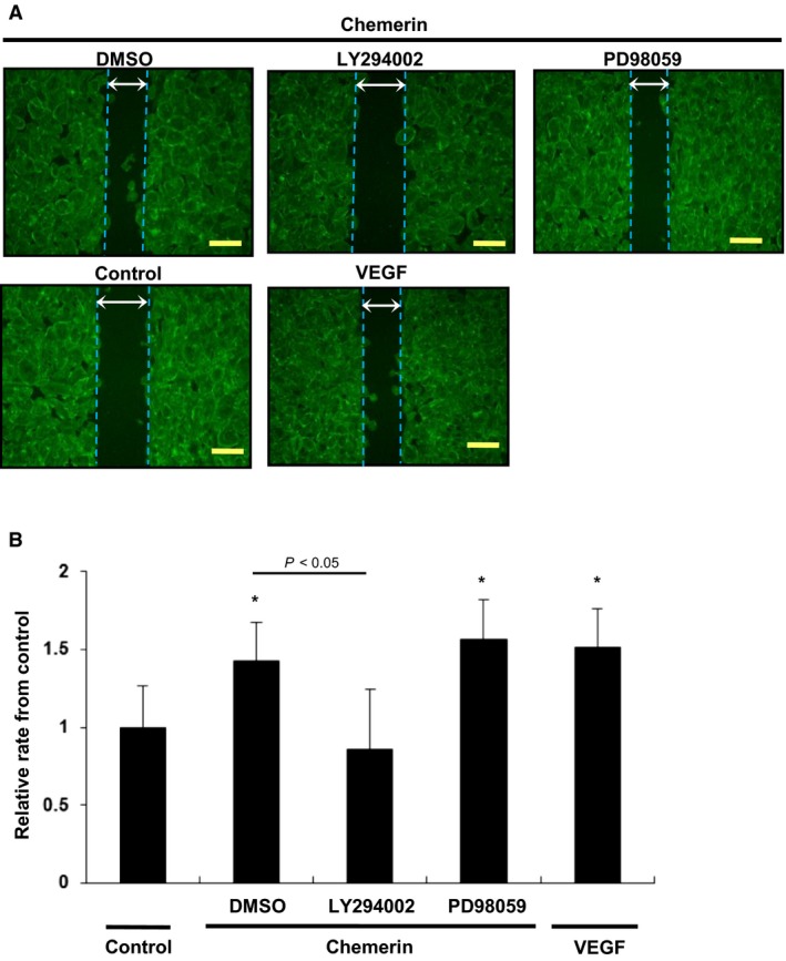
Figure 4.

Migration of HUVECs stimulated by chemerin assessed by cell scratch assay. (A) Modified wound healing assay was performed with chemerin (10 nmol/L) or VEGF (5 nmol/L) as chemoattractant. Chemerin stimulation (10 nmol/L) was conducted for the indicated time. After preincubation with LY294002 (1 μmol/L) or PD98059 (5 μmol/L), cells were incubated with chemerin (10 nmol/L). (B) The change rate of width between the leading edge covered by cells before and after 8 h of incubation was quantified. Seven random wounds per each well were quantified. All assays were performed in triplicate. *P < 0.05 versus serum‐free control.
