
Figure 3.
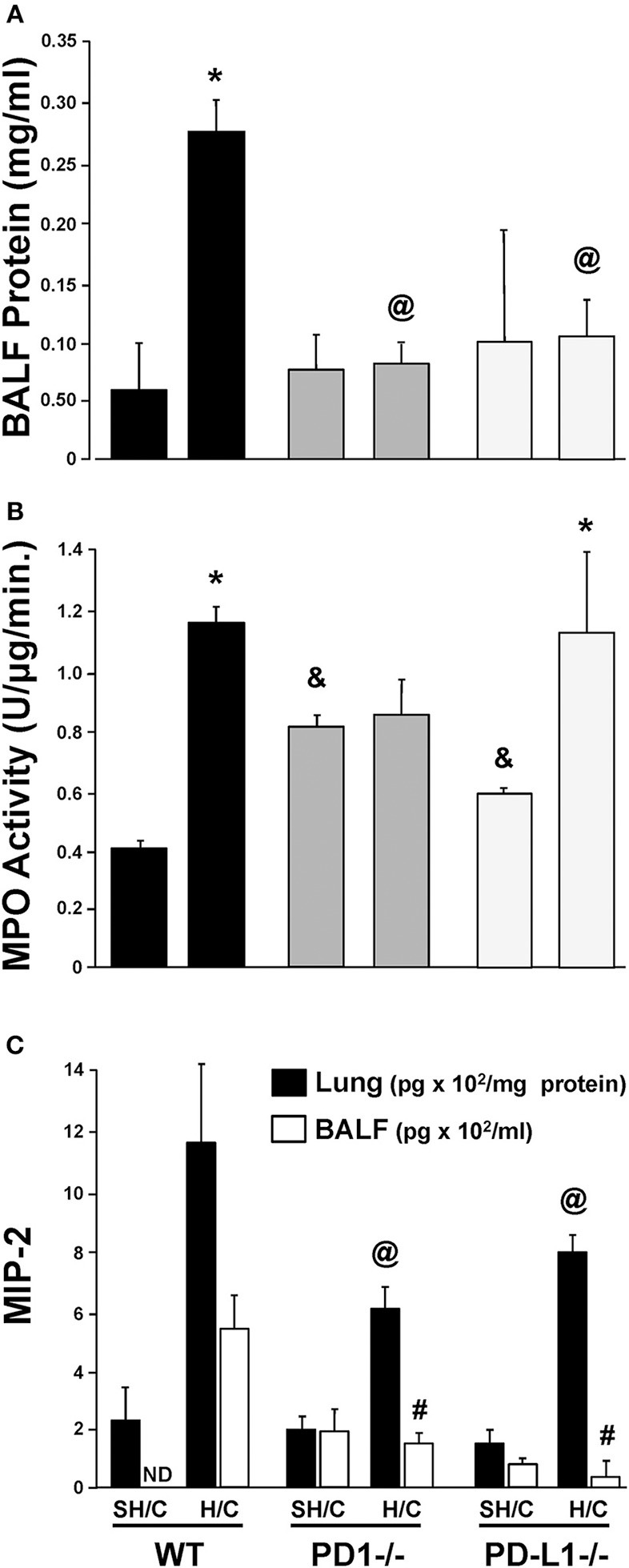
Concentration of protein in bronchial alveolar lavage fluid (BALF) as an assessment of lung injury in SH/C and H/C in PD-1−/−, PD-L1−/− and WT Control mice (A). N = 5–7/group; @p < 0.05 vs. Control H/C; *p < 0.05 vs. respective SH/C. Lung tissue myeloperoxidase activity (MPO) in the lungs of PD-1−/−, PD-L1−/− and WT Control mice following SH/C and H/C (B). N = 3–5/group; *p < 0.05 vs. respective SH/C; &p < 0.05 vs. Control SH/C; @p < 0.05 vs. Control H/C. Neutrophil chemotactic protein, MIP-2, in lung tissue and BALF in those same groups (C). Mean ± SEM, N = 5–7/group; @p < 0.05 vs. Control H/C (lung protein); #p < 0.05 vs. Control H/C (BALF). One-way ANOVA and a Tukey's multiple comparisons test.