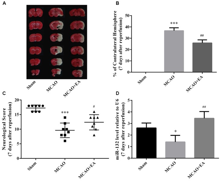
FIGURE 1.

EA treatment alleviated brain injury and up-regulated miR-132 expression in ischemic penumbra after stroke. (A) 2, 3, and 5-triphenyltetrazolium chloride (TTC) staining was used to measure infarct volume in coronal brain sections from Sham, MCAO, and MCAO+EA-treated rats at 7 days after reperfusion. (B) Bar graph showed the infarct volume in coronal brain sections in each group. (C) Neurological score was used to assess recovery of neural function by EA treatment after stroke. (D) Bar graph showed the relative expression of miR-132 in the ischemic penumbra in each group. Data are expressed as means ± SEM (n = 8 per group); ∗p < 0.05 vs. Sham, ∗∗∗p < 0.001 vs. Sham; #p < 0.05 vs. MCAO, ##p < 0.01 vs. MCAO.
