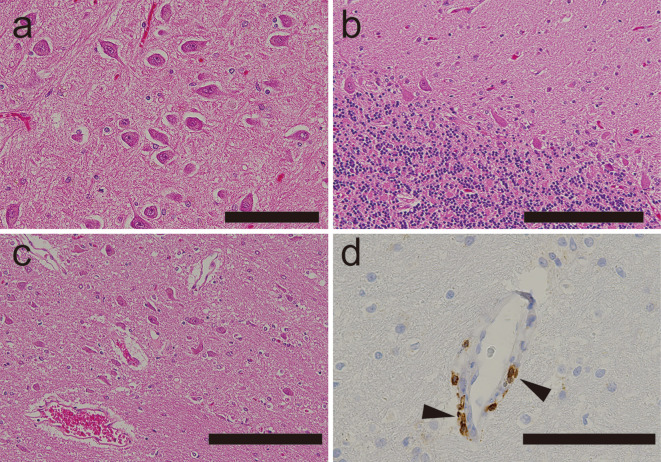
Figure 4.
A neuropathological examination of a patient’s brain in a case of progressive encephalomyelitis with rigidity and myoclonus that resolved after thymectomy. a-c) Hippocampus (a), cerebellum (b), and brainstem (c) showing no evidence of microglial nodules or perivascular cuffing (Hematoxylin and Eosin staining). d) Immunohistochemistry revealing occasional CD3-positive cells around vessels in the pons (arrowheads) and other brainstem structures. Scale bar: a, d 100 μm; b, c 200 μm

