Figure 2.
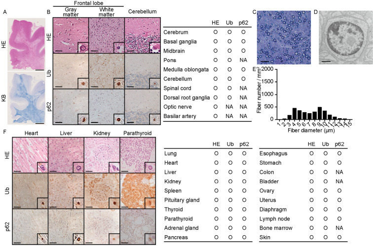
Pathological autopsy findings of the proband. (A) Low-magnification image of the frontal lobe. Hematoxylin and Eosin (H&E) staining (upper) and Klüver-Barrera (KB, lower) staining results are shown. Scale bar=5 mm. (B) Representative pathological findings for the central and peripheral nervous systems. H&E staining and immunohistochemical staining results for ubiquitin and p62 are shown. The table provides a summary of the pathological findings. Insets are the magnified views of nuclei with inclusion bodies. Scale bar=50 µm. (C-E) Pathological findings of the distal sural nerve. Transverse semi-thin sections show the loss of myelinated fibers (C, E). (D) EM image of a Schwann cell nucleus with an inclusion body. Scale bar=20 µm (C), 1 µm (D). (F) Representative pathological findings in the visceral organs. H&E staining and immunohistochemical staining results for ubiquitin and p62 are shown. Insets are the magnified views of nuclei with inclusion bodies. The densities of the nuclei with Ub-positive inclusions were approximately 10/mm2, 75/mm2, 120/mm2, and 160/mm2 in the heart, liver, kidney, and parathyroid, respectively. Scale bar=50 µm. The table provides a summary of the pathological findings. NA: not available

