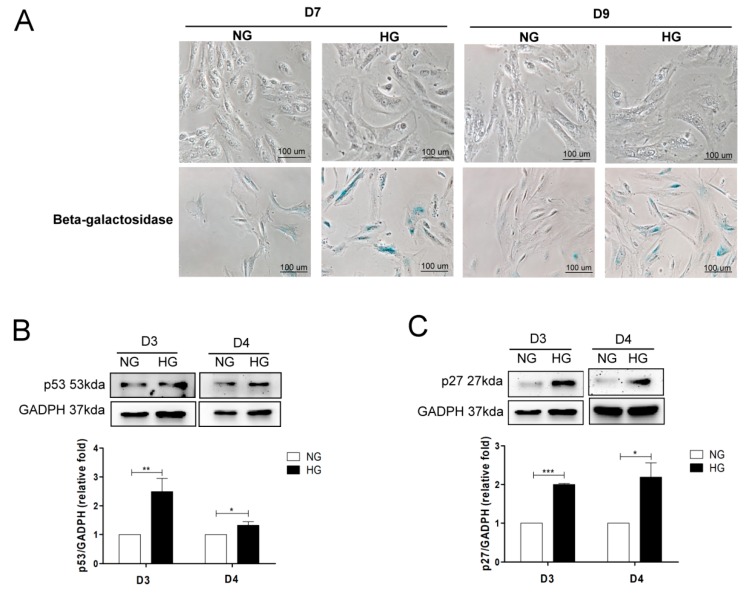
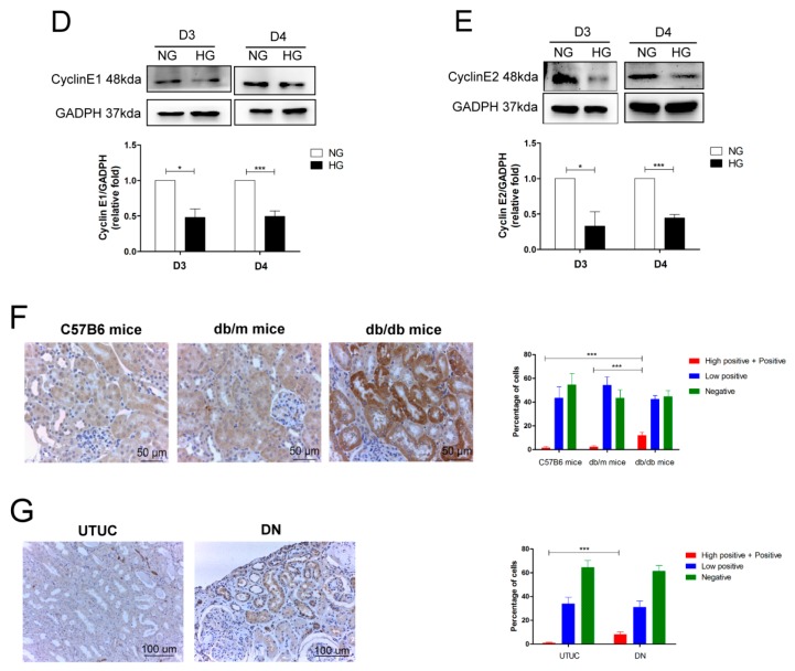
Figure 1.
High glucose (HG) induces cell senescence in proximal tubular epithelial cells (PTECs). (A) The effect of HG on morphological changes and senescence-associated β-galactosidase (SAβ-Gal) staining of human PTECs. PTECs were incubated under normal glucose (NG, 6.2 mM) and HG (30 mM) conditions for seven days and nine days. Cell senescence was assessed using SAβ-Gal staining. HG increased p53 (B) (D3: n = 6, D4: n = 4) and p27 (C) (D3: n = 3, D4: n = 3), and decreased cyclin E1 (D) (D3: n = 3, D4: n = 4) and cyclin E2 (E) (D3: n = 3, D4: n = 4) protein expression in human PTECs after three and four days of treatment. Protein levels were assessed by western blot. The expression of p53 in the proximal tubule of kidneys of mice (F) and humans (G). The kidney sections of C57BL/6 mice, non-diabetic db/m mice, and diabetic db/db mice, and human donors (upper tract urothelial carcinoma, UTUC with normal kidney function and normal glomerulus and proximal tubule) and patients with diabetic nephropathy (DN) were stained with p53 (brown). The images quantification was performed using the IHC Profiler Plugin of ImageJ Software. The bar graph represents the mean ± S.E.M. * p < 0.05, ** p < 0.01, *** p < 0.001 by Student’s t test.


