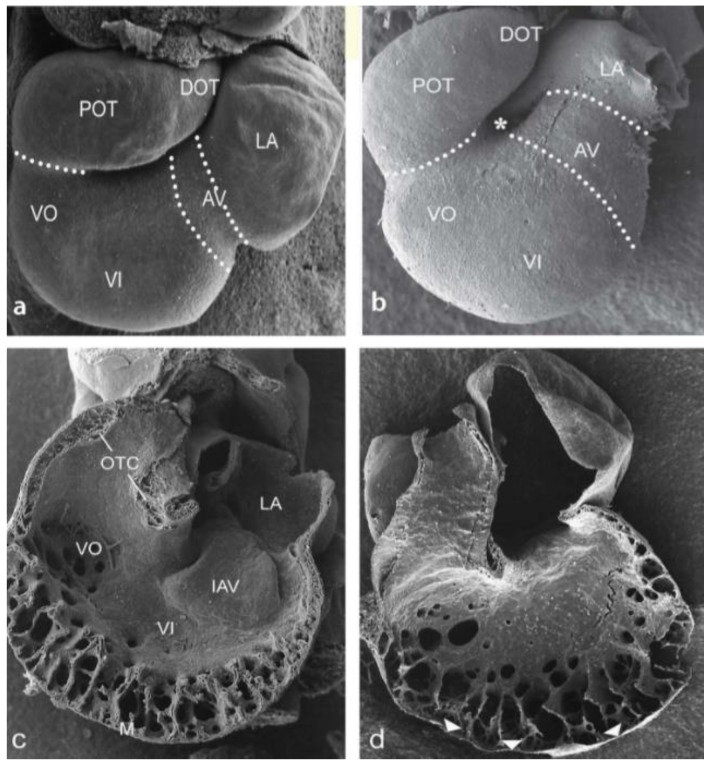
Figure 1.
Cardiac looping in normal and ligated embryos HH20. Scanning electron micrograph (SEM) of ventral views. (a) Normal embryo with cardiac segments indicated. (b) Ligated embryo. The retarded looping resembles that of a HH17 embryo with an open inner curvature (*). The AV canal is relatively long. (c,d) Interior view of dorsal heart halves. (c) The inferior AV cushion and the OFT cushions are well developed, ventricular trabeculations have formed. (d) AV and OFT cushions are non-existent, spongy trabeculations and the compact myocardium is thin (arrowheads). AV: Atrioventricular groove, DOT: distal OFT, IAV inferior AV cushion, LA: left part of atrium, M: compact myocardium, OTC: OFT cushions, POT: proximal OFT, VI ventricular inlet, VO: ventricular outlet, * inner curvature, arrowheads: thin compact myocardium.

