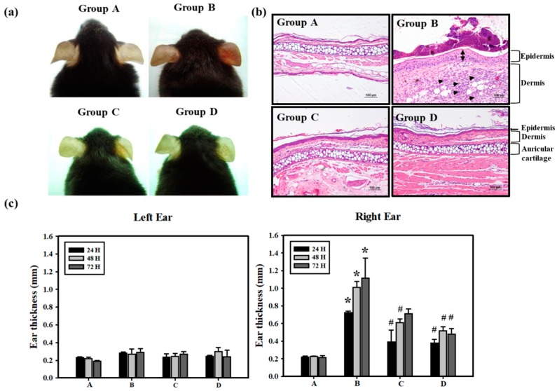
Figure 2.
Protective effects of PT on Cr(VI)-induced ACD mice. (a) Swelling and erythema were documented by photography at 48 h. (b) H&E histology of mice ear skin after the elicitation test with Cr(VI) showed ear swelling and inflammatory cell infiltration at 72 h. In the dermis, the increased number of inflammatory cells, neutrophils, was observed (arrowhead) in K2Cr2O7 treatment (Group B). In the epidermis, Group B showed the stratum corneum thickening (bi-arrowhead), but NAC (Group C) or PT (Group D) treatment significantly suppressed Cr(VI)-induced increases in ear thickness. Also, no differences in the auricular cartilage were observed (scale bar representing 100 μM). (c) ACD reactions were quantified by measuring the ear thickness at 24, 48 and 72 h after the elicitation test (n = 5). The time course measurement of Left (vehicle) and Right (epicutaneous) ear thickness. (A: Control; B: K2Cr2O7; C: NAC; D: PT) (* p <0.05 versus control group; # p <0.05 versus K2Cr2O7 group).

