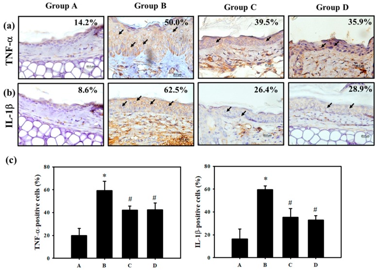
Figure 3.
PT protects against pro-inflammatory cytokines in mice with ACD induced by Cr(VI). Skin biopsy from the Control, K2Cr2O7, NAC and PT groups shows immunohistochemical staining for analysis of (a) TNF-α. (b) IL-1β- positive cells (brown) in the epidermis (scale bar representing 20 μM). (c) The quantification of TNF-α and IL-1β-positive cells was determined using HistoQuest software (Version 4.0.4.0158, TissueGnostics). (n = 3, * p <0.05 versus control group; # p <0.05 versus K2Cr2O7 group).

