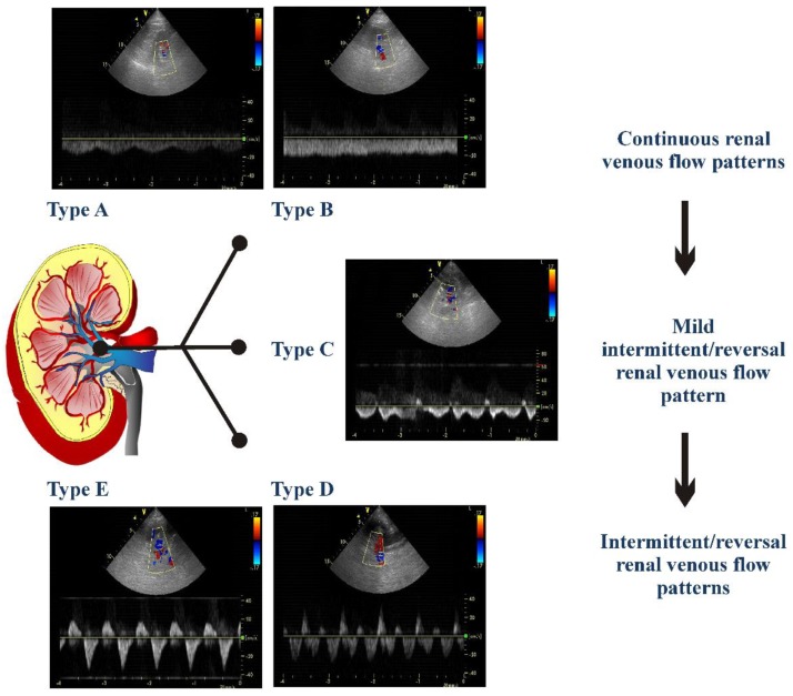
Figure 1.
The different detectable renal venous patterns are presented. Pattern A was a flow pattern with normal velocity decrease of presystolic flow and biphasic pattern without interruption of telediastolic flow; Pattern B a continuous flow pattern with minimal fluctuations; Pattern C a pattern showing a short telediastolic interruption of forward flow or a short telediastolic reversal flow; Pattern D a flow characterized by a biphasic interruption or reversal flow during the same cardiac cycle; Pattern E a flow characterized by one forward and one reversal wave flow, i.e., monophasic intermittent pattern.

