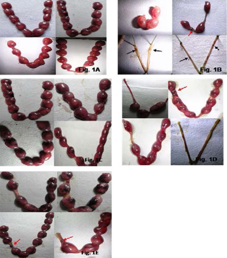
Fig. 1.
Implantation sites in the uterus of the female mice impregnated with (A.) the control male mice. Note the normal range of the numbers of implanted embryos in the left and right uteri. (B.) the male mice administered with 500mg/kgBW/day of MTZ for 28 days. Note the post-implantation loss indicated by the least number of implanted embryos in the uterus, the dead implant (red arrow) and the resorption sites indicated by scars (black arrow) observed in the uterus devoid of implanted embryos. (C.) the male mice administered with 200mg/kgBW/day of the fruit extract of TT for 28 days. Note the normal range of the numbers of implanted embryos in the left and right uteri. (D.) the male mice co-administered with MTZ (500mg/kgBW/day) and the fruit extract of TT (100mg/kgBW/day) for 28 days. Note the post-implantation loss indicated by the dead implant (red arrow), the partial recovery in the number of implanted embryos as the left and right uteri of one female was still devoid of implanted embryos. (E.) the male mice in Gr. VIII, co-administered with MTZ (500mg/kgBW/day) and the fruit extract of TT (200mg/kgBW/day) for 28 days. Note the recovery in the number of implanted embryos in the uterus. Also note a dead implant (red arrow) in the left uteri of two females.

