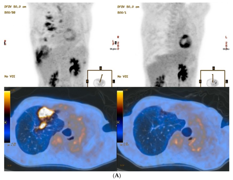
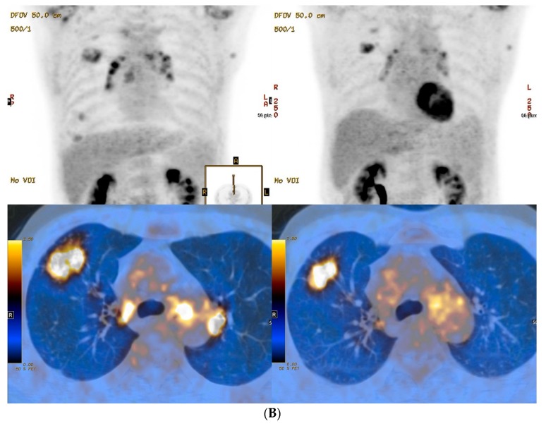
Figure 1.
PET/CT images of lungs from two MDR-TB patients. Baseline (left column) and 12 months follow-up (right column) maximum-intensity projection and transverse fusion PET/CT images of patient 2 (A) and patient 4 (B). (A) Images of patient 2 show multiple hypermetabolic lesions in the right lung at baseline that disappeared in follow-up images despite persistent disease. (B) Images of patient 4 show multiple hypermetabolic lesions in the right lung at baseline that improved but were persistent in follow-up images irrespective of negative culture conversion and treatment success. Total metabolic lung volume (92.3 cm3) and total lung glycolysis (307.3 cm3) in patient 2 using a baseline PET/CT were much higher than for patient 4 (4.6 cm3 and 14.1 cm3, respectively).


