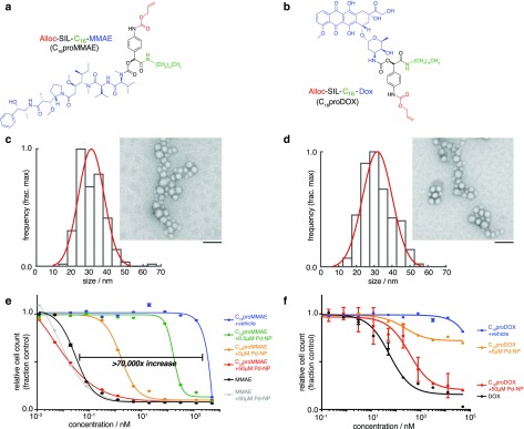
Figure 2.
Caged MMAE and DOX encapsulate into nanoparticles and are selectively cytotoxic in the presence of the Pd-NP bioorthogonal trigger. (a–b) Chemical structure of caged MMAE (a) and DOX (b), color-coded according to the scheme in Figure 1. (c–d) TEM imaging of C16proMMAE (c) and C16proDOX (d) encapsulated in a formulation of PLGA-PEG polymeric micelles. Particle diameters were quantified according to their distribution (black bars) and Gaussian fit (red curve), with representative images shown at right (scale bar, 100 nm). (e–f) Cytotoxicity was determined for caged MMAE (e) and DOX (f) at the indicated concentration (x-axis), in the presence of varying amounts of Pd-NP bioorthogonal trigger, over 72 h treatment using HT1080 fibrosarcoma cells (n = 2, data are means ± s.e.m.). Parent noncaged compounds were also tested as controls (black curves).

