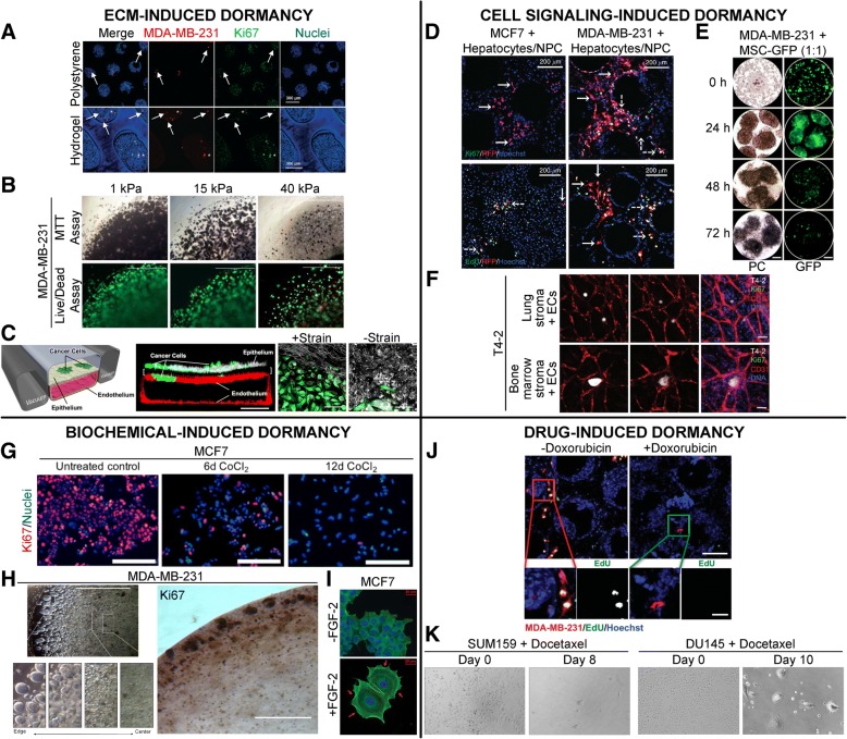
Fig. 3.
Engineered, in vitro models for induction of cancer dormancy. Representative examples of in vitro dormancy models classified by induction mode. a MDA-MB-231-RFP cells co-cultured with primary human hepatocytes and non-parenchymal cells (NPCs) within a hepatic microphysiological system either seeded on a polystyrene surface or encapsulated within a PEG-peptide hydrogel matrix and imaged on day 15. Arrows: dormant cells, asterisks: proliferative cells. Scale bar = 300 μm. (Adapted from [105]). Copyright 2017, RSC. b MDA-MB-231 cells cultured within Col-Tgel hydrogels demonstrate an increased dormancy signature characterized by reduced MTT staining, reduced cell death and lower cell density. Green: calcein AM, red: ethidium homodimer. Scale bar = 1000 μm. (Adapted from [89]). Copyright 2017, Springer Nature. c GFP expressing, non-small-cell lung cancer cells (NSCLC) cultured with alveolar epithelial cells and lung microvascular endothelial cells within a microfabricated lung-on-a-chip device for 2 weeks to investigate the role of physiological breathing motions on the growth/dormancy of cancer cells. Red: VE-cadherin, white: ZO-1 tight junctions, Scale bar = 200 μm (center), 50 μm (right). (Adapted from [104]). Copyright 2017, Elsevier. d RFP expressing breast cancer cells cultured with hepatocytes and NPCs within a liver microphysiological system for 2 weeks and fluorescently labeled for Ki67 or EdU (green) and nuclei (blue). Scale bar = 200 μm. Solid white arrows: dormant cells, dashed white arrows: proliferative cells. (Adapted from [119]). Copyright 2014, NPG. e MDA-MB-231 cells cultured with GFP expressing MSCs and imaged under phase contrast (PC) and green fluorescence (GFP) at varying time points are observed to cannibalize MSCs within 3D spheroids and enter dormancy, leading to reduced GFP signal intensity. Scale bar = 100 μm. (Adapted from [117]). Copyright 2016, NAS. f HMT-3522-T4-2 breast cancer cells cultured with lung/bone marrow stromal cells and endothelial cells remain as dormant clusters through day 17 with low proliferation. Scale bar = 100 μm. (Adapted from [42]). Copyright 2013, NPG. g MCF7 cells treated with 300 μM CoCl2 undergo hypoxia and enter dormancy with low proliferation. Scale bar = 200 μm. (Adapted from [129]). Copyright 2018, Springer Nature. h MDA-MB-231 cells within Col-Tgel hydrogels exhibit reduced proliferation and cluster size with increasing distance from the hydrogel edge due to a hypoxia gradient. Scale bar = 100 μm. (Adapted from [128]). Copyright 2014, PloS. i MCF7 cells seeded on a fibronectin-coated substrate and treated with FGF-2 undergo a dormancy phenotype with cortical actin redistribution around the perimeter of the cytoplasm (red arrows). Scale bar = 20 μm. (Adapted from [137]). Copyright 2009, Springer. j MDA-MB-231 cells in an engineered liver niche treated with doxorubicin exhibit reduced proliferation compared to the control group. Scale bar = 200 μm (top), 50 μm (bottom). (Adapted from [81]). Copyright 2013, ASBMB. k Breast and prostate cancer cells treated with docetaxel exhibit residual tumor cells with dormancy signatures. (Adapted from [148]). Copyright 2014, PloS

