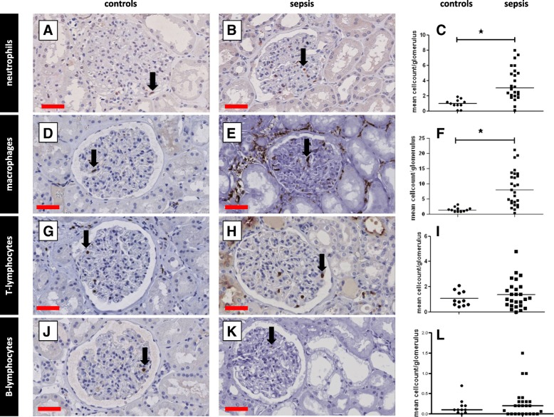
Fig. 1.
Leukocyte infiltration in the glomeruli. Leukocyte subsets were immunohistochemically detected in kidney biopsy samples using specific antibodies (Additional file 2: Table S3: Primary antibodies) and scored according to Table 2. Leukocyte subsets were counted and divided by the number of glomeruli per patient. Neutrophils (a, b), macrophages (d, e), T lymphocytes (g, h), and B lymphocytes (j, k) were observed in the glomeruli in kidney tissue from control patients (a, d, g, j) and patients with sepsis (b, e, h, k), and mean leukocyte counts were determined (c, f, i, l). Black arrows show positively stained leukocytes of various subsets. *Statistically significant. Black lines are medians (c, f, i, l). Red scale bar = 50 μm

