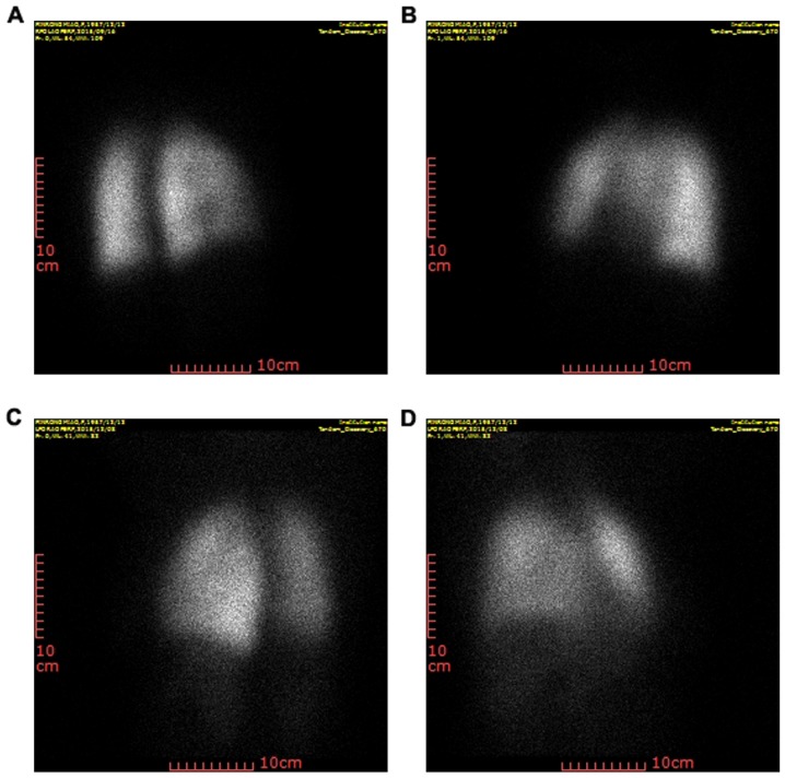
Figure 1.
Single-photon emission computed tomography images of patients. (A and B) Representative images of non-small cell lung cancer patients without RP prior to radiotherapy. (C and D) Images obtained after stereotactic body radiation therapy, displaying RP with ground-glass opacity changes and irregular enhancement in the lung (scale bars, 10 cm). All images were obtained from different patients. RP, radiation pneumonitis.

