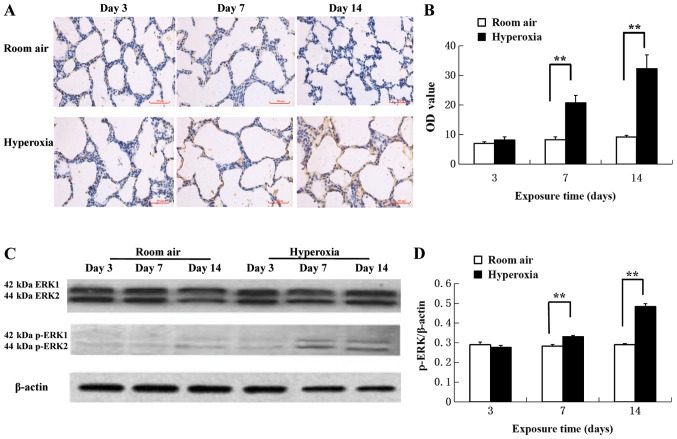
Figure 3.
Phosphorylation of ERK1/2 in lung tissue samples. (A) Increased p-ERK1/2 antigen levels were observed in the cytoplasm and nucleus of pulmonary epithelial and mesenchymal cells by immunohistochemistry using the peroxidase-conjugated streptavidin method. Scale bar, 50 µm. (B) Protein levels of p-ERK were detected by immunohistochemistry. p-ERK levels increased in the hyperoxia group starting from postnatal day 7 compared with the room air control group (n=5). (C) Western blot analysis of lung tissue samples detecting p-ERK1/2, ERK1/2 and β-actin. (D) Western blot quantification suggested that the intergroup difference was more significant at postnatal day 7 and 14 for p-ERK levels (n=5). **P<0.01. ERK, extracellular signal-regulated kinase; p, phosphorylation.

