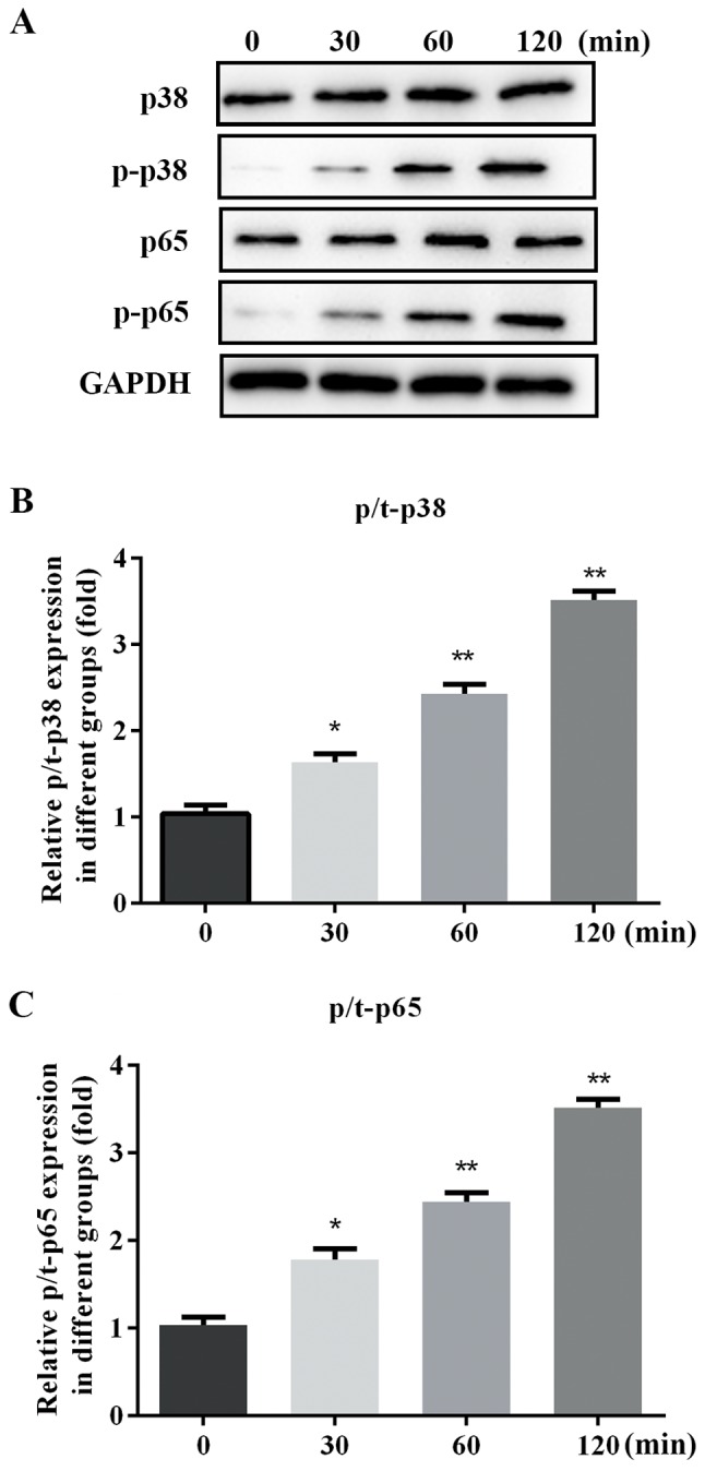
Figure 3.

Levels of p38, p-p38, p65 and p-p65 protein. HK-2 cells were incubated with 10 µg/ml surfactant protein A for 30, 60 or 120 min. (A) Representative western blot bands for p38, p-p38, p65 and p-p65. Relative protein levels of (B) p/t-p38 and (C) p/t-p65. GAPDH was used as an internal reference. *P<0.05, **P<0.01 vs. 0 min. p-p38, phosphorylated p38; p-p65, phosphorylated p65.
