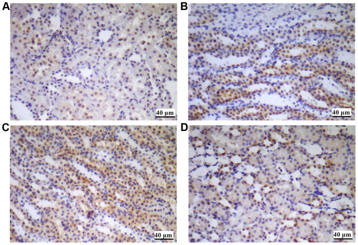
Figure 5.
HMGB1 immunohistochemical staining of renal tissues. (A) Control. (B) CH group. (C) IH group. (D) IHH group. The expression of HMGB1 was identified primarily in the nucleus of the renal tubular epithelial cells, as demonstrated in (A). The expression and translocation of HMGB1 was increased in the (B) CH group, (C) IH group and (D) IHH group compared with the control. IHH, intermittent hypoxia with hypercapnia; IH, intermittent hypoxia; CH, continuous hypoxia; HMGB1, high mobility group box 1 protein.

