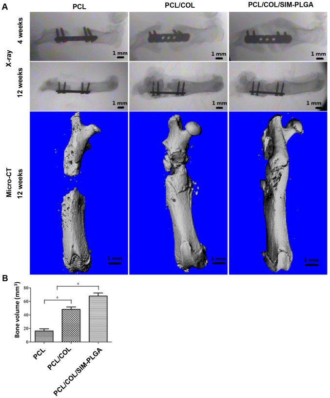
Figure 7.
Analysis of bone regeneration of the femur defect with the implanted scaffolds using X-ray and micro-CT. (A) Plain X-ray images of the femur defects at 4 and 12 weeks after transplantation with PCL, PCL/COL or PCL/COL/SIM-PLGA. Micro-CT scan of the femur bone defect at 3 months after surgery. (B) Bone volume. The rats in the PCL/COL/SIM-PLGA group exhibited improved bone repair when compared with the other groups. *P<0.05, as indicated. PLGA, poly(lactic-co-glycolic acid); SIM, simvastatin; COL, collagen; PCL, poly(ε-caprolactone); CT, computed tomography.

