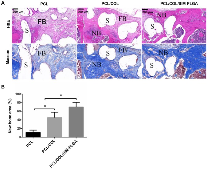
Figure 8.
Analysis of bone regeneration of the femur defect by using HE and Masson staining. (A) HE and Masson stained tissue sections of bone regeneration in the defect areas, implanted with PCL, PCL/COL or PCL/COL/SIM-PLGA constructs, at 3 months after surgery. (B) The SIM-loaded scaffold demonstrated the most robust osteogenic activity, and the majority of the defect area in the SIM-loaded scaffold group was filled with eosin-stained newly formed bone tissue. *P<0.05, as indicated. S, scaffolds; NB, newly formed bone; FB, fibrosis; PLGA, poly(lactic-co-glycolic acid); SIM, simvastatin; COL, collagen; PCL, poly(ε-caprolactone); HE, hematoxylin and eosin.

