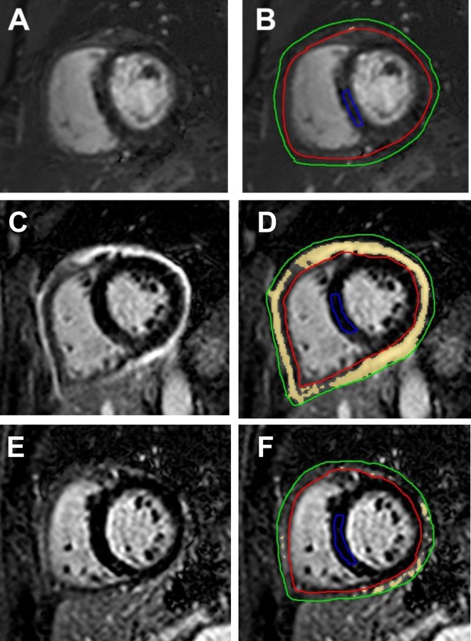
Figure 5.
Delayed hyperenhancement (DHE) images from patients with RP. Panels A and B are DHE images from a 47-year-old female patient with RP who had minimal pericardial DHE at presentation. Panels C and D show severe pericardial DHE in a 61-year-old female patient with RP diagnosed as having an ongoing recurrence at presentation, while panels E and F are images from the same patient showing improved DHE post-treatment. Panels A, C and E show images before contouring, and the pericardium is bright from intense DHE in panel C. Postcontouring (B, D and F), quantitative signal >6 SD above normal myocardium is shown as yellow. On these short-axis images, the pericardium has been outlined between the green and red tracings, and normal septal myocardium has been outlined as a reference region (blue tracing). While DHE images show very low quantitative DHE (quantitative DHE=2 cm3) in panel B, panel D shows high-quantitative DHE (quantitative DHE=142 cm3). In comparison with panel D, panel F shows improved DHE (quantitative DHE=34 cm3) after 200 days of anti-inflammatory therapy. RP, recurrent pericarditis.

