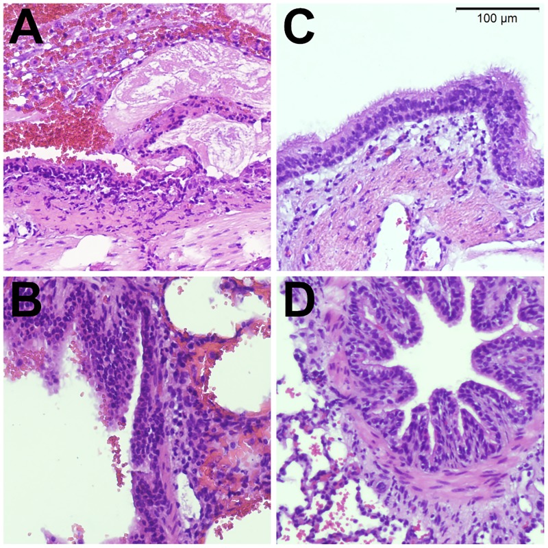
Fig 2. Histology.
Representative histology images taken from a lung following experimental endobronchial aspiration (A,B). Large airways show extensive epithelial damage with necrosis and endobronchial hemorrhage (A). Distal airways have intact epithelium and show signs of hemorrhage, extending from more proximal sites into the alveolar space (B). Control lungs (C, D) show intact epithelium in large (C) and small (D) airways and focal intraparenchymal hemorrhage after ex-vivo perfusion (D).

