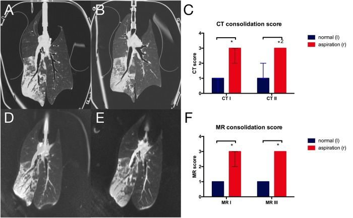
Fig 5. CT and MRI morphologic assessment of lung injury.
Central coronal images of A first and B second CT scan as well as D first and E third MRI scan showing infiltrates after aspiration in the right lower lobe. Morphologic scoring of lung alterations based on C CT scans and F MRI scans. (*p = 0.016, *2p = 0.031; data from reader 1).

