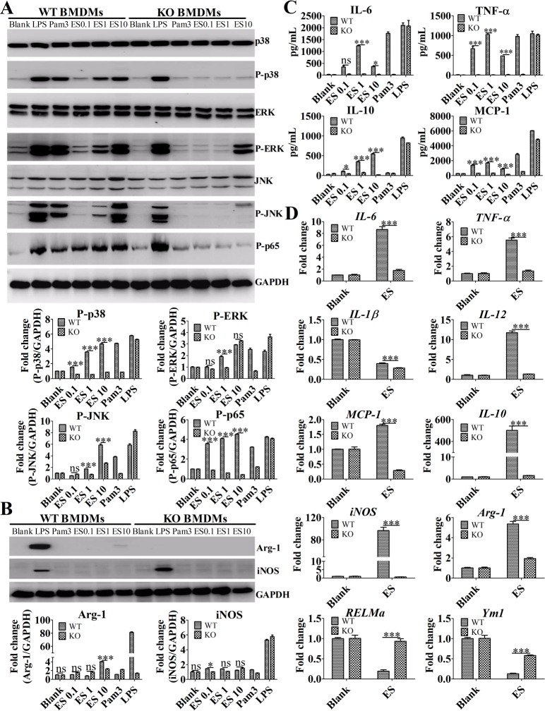
Fig 3. ES promoted M2b polarization by activating the MAPK/NF-κB signaling pathway in a TLR2-dependent manner.
(A) Immunoblot of P-p38, P-p65, P-ERK1/2, and P-JNK in the lysates of BMDMs after stimulation for 30 min. Graphical representations of the band intensities are shown in the pictures below. Expression of P-p38, P-p65, P-ERK1/2, and P-JNK was normalized to GAPDH expression. (B) The expression of iNOS and Arg-1 protein in the lysates of BMDMs following stimulation for 24 h was assessed by Western blot. Graphical representations of the band intensities are shown in the pictures below. Expression of iNOS and Arg-1 was also normalized to GAPDH expression. (C) The levels of IL-6, MCP-1, TNF-α, and IL-10 in the supernatants of BMDMs were measured by ELISA. (D) The levels of IL-6, TNF-a, IL-10, IL-12, IL-1β, MCP-1, iNOS, Arg-1, RELMa, and Ym1 mRNA in the purified liver macrophages from wild-type and TLR2 KO mice after stimulation for 4 h were analyzed by RT-qPCR. The data were expressed as the mean ± SEM, and are the results of a representative experiment out of three independent experiments and analyzed by two-way ANOVA. *P < 0.05; **P < 0.01; ***P < 0.001; ns, not significant.

