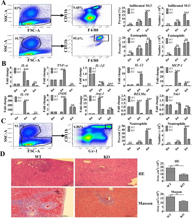
Fig 5. TLR2 promotes M2 polarization and liver pathology in murine schistosomiasis.
(A) The absolute numbers of eosinophils and infiltrated macrophages (MØ) in the liver and percentages of eosinophils and infiltrated macrophages (MØ) of the total liver leukocytes were determined by flow cytometry. Representative flow cytometry analysis of infiltrated macrophages (SSClowCD11b+ F4/80+ cells) and eosinophils (SSChighCD11b+F4/80+ cells) among liver leukocytes from one wild-type-infected mouse at 4 weeks post-infection. The data are presented as the mean ± SEM obtained from 10 mice per group and analyzed by two-way ANOVA. (B) The levels of IL-6, MCP-1, IL-10, IL-12, TNF-a, IL-1β, Arg-1, RELMa, Ym1, and iNOS mRNA in purified macrophages were analyzed by real-time PCR. The data are the results of a representative experiment out of three independent experiments and analyzed by two-way ANOVA. (C) The absolute number of neutrophils in the liver and percentage of neutrophils among the total liver leukocytes was determined by flow cytometry. Representative flow cytometric analysis of neutrophils (CD11b+Gr-1+ cells) among liver leukocytes from one wild-type-infected mouse at 4 weeks post-infection. Data are presented as the mean ± SEM obtained from 10 mice per group and analyzed by two-way ANOVA. (D) The area of liver granulomas and fibrosis developed following S. japonicum infection were detected at 6 weeks post-infection. Slide sections from infected mice were examined under a light microscope, and images were captured and analyzed. Representative granuloma in the liver sections of TLR2-/- and wild-type mice (hematoxylin-eosin [upper panel] and Masson’s trichrome [lower panel]). Original magnification: × 100. Data are presented as the mean ± SEM and were analyzed by Student’s t-test. *P < 0.05; **P < 0.01; ***P < 0.001; ns, not significant.

