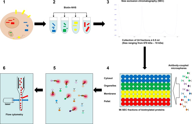
Fig 1. Microsphere affinity proteomics (MAP).
1–2: Proteins from different subcellular compartments were lysed and labeled with amine-reactive biotin (Biotin-NHS). 3: The biotinylated proteins were separated using a size exclusion chromatography (SEC) column (Superdex 200). 4: A mixture of color-coded microspheres with antibodies to cellular proteins was added to all SEC fractions and the microspheres rotated overnight at 4oC. 5: Microspheres were washed and then labeled with R-Phycoerythrin-conjugated streptavidin (SA-PE, red flashing circles). 6: Finally, the microspheres were analyzed by flow cytometry.

