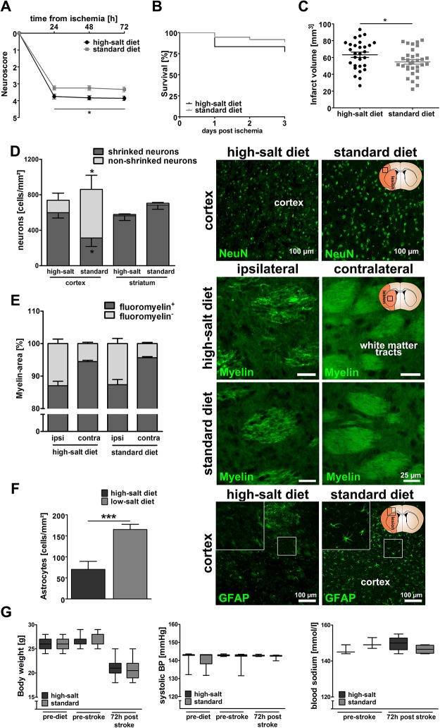
Fig 1. Sodium chloride aggravates functional and structural outcomes after ischemic stroke.
(A) Neurological deficit score assessment showed significantly worse functional outcomes following high-salt diet (p<0.05, 2way ANOVA, n = 36 per group). (B) Mortality rates did not significantly differ between both groups (p = 0.19, Log-rank (Mantel-Cox) Test, n = 36 per group). (C) Infarct volumes were significantly increased after high-salt diet (64.2 mm2 ± 3.1 mm2 vs. 54.7 mm2 ± 2.7 mm2, p<0.05, t-test, n = 28 to 31 per group). (D) Immunohistochemical analyses revealed a significant increase in the number of shrinked cortical neurons following high-salt diet, and vice versa a reduced number of intact cortical neurons (p<0.05, t-test, n = 8 to 11 per group). NeuN-staining was used to visualize intact and shrinked neurons. (E) The extent of fluoromyelin-positive myelin areas was similar in both groups (p = 0.88, t-test, n = 20 to 22 per group) Fluoromyelin-staining was employed to visualize white matter tracts in the ispilateral and contralateral hemisphere of both groups. (F) High-salt diet induced a significant loss of GFAP+-astrocytes in the periinfarct region (p<0.001, t-test, n = 10 to 11 per group). (G) Body weight and systolic blood pressure were not significantly influenced (p = 0.95 and p = 0.83 2way ANOVA, n = 36 per group). Blood sodium levels did not significantly differ between both groups prior to MCAO and three days after MCAO (p = 0.19 and p = 0.18, t-test, n = 3 to 6 per group).

