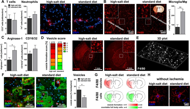
Fig 2. Sodium chloride induces vesicle formation of microglia/macrophages.
(A) The total number of infiltrating 7/4+-neutrophils and CD3+-T cells was not affected (p = 0.16 and p = 0.61, t-test, n = 15 per group (7/4) and n = 5 per group (CD3)). (B) The number of F4/80+-microglia/macrophages was significantly reduced after high-salt diet (p<0.01, t-test, n = 15 per group). (C) Arginase-1-stainings illustrated a significant decrease of Arginase-1+-anti-inflammatory cells following high-salt diet (p<0.05, t-test, n = 15 per group). High-salt diet did not influence the number of CD16/32+-pro-inflammatory cells (p = 0.28, t-test, n = 16 per group). (D) Quantification of vesicle formation among F4/80+-cells according to a previously defined score revealed increased vesicle formation following high-salt diet (score 0: No vesicles, 1: few vesicles, 2: occasional vesicle formation, 3: vesicle formation covering at least half of the striatum, 4 extreme vesicle formation, n = 14 per group). (E) Three-dimensional plot illustrating the distribution of vesicles and cytoplasmic expansions within the primary motor cortex. (F) The total area occupied by vesicles was significantly larger after high-salt diet (p<0.05, t-test, n = 16 per group). (G) Heat maps illustrate the distribution of F4/80+-vesicles (red) and intact F4/80+-microglia/macrophages (green) in both experimental groups. (H) In sham operated animals, high-salt diet did not induce microglial vesicle formation.

