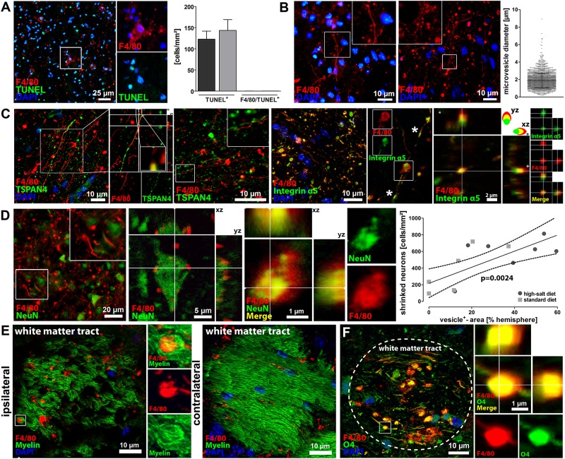
Fig 3. Characterization of F4/80+-vesicles as migrasomes dispatching neuronal cytoplasm.
(A) The previously identified F4/80+-vesicles did not interact with TUNEL, thus rebutting the hypothesis that these vesicles were merely apoptotic blebs. (B) We measured the diameter of > 2300 vesicles and calculated a mean diameter of 1,87 ± 0,18 μm, which is in the range of the migrasome. (C) Immunohistochemistry and confocal microscopy demonstrated that the F4/80+-vesicles expressed the characteristic migrasome-markers TSPAN4 and integrin α5, which provides evidence that the identified vesicles belong to the migrasome. Furthermore, our findings confirm the previously in vitro described polarized expression pattern of TSPAN4 and the migrasomal enriched expression of integrin a5 with occasional small integrin-positive puncta (asterisks), which have been shown to be sites of migrasome formation in vitro. (D) Intact and shrinked NeuN+-neurons were found in close vicinity to the migrasome, and confocal microscopy visualized neuronal fragments within the migrasome, thus suggesting that the migrasome incorporates the cytosol of surrounding neurons. Correlation analyses moreover revealed a positive correlation between vesicle occurrence and neuronal loss (p<0.01, linear regression, n = 7 per group). (E) Immunohistochemistry and confocal microscopy demonstrated that the migrasome infiltrates Fluoromyelin+-white matter tracts. (F) Immunohistochemistry and confocal microscopy also demonstrated that the migrasome interacts with O4+-oligodendrocytes.

