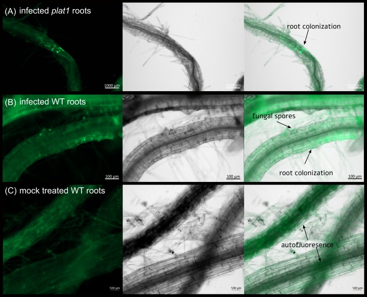
Fig 4. Root colonisation of Arabidopsis WT and plat1 by P. indica at 7 dpi.
Colonisation of Arabidopsis plat1 (A) and WT roots (B) was imaged using fluorescence microscopy at 7 dpi. Uncolonised roots are shown at (C). The first pictures show the autofluorescence of the root and GFP (green fluorescent protein) fluorescence of P. indica. The second pictures show the bright field image. The last pictures show the overlay of all images for each row.

