Figure 1.

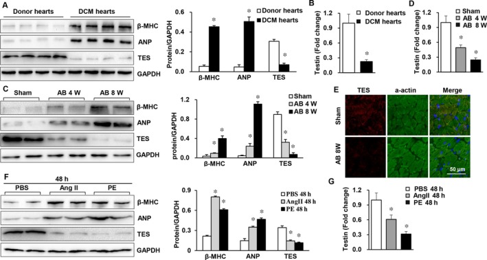
TES expression was suppressed in failing hearts and in murine cardiac hypertrophy models. A, Western blot analysis of markers of hypertrophy (ANP and β‐MHC) and TES protein levels in healthy donors and donors with dilated cardiomyopathy (n = 4 in each group). B, Real‐time PCR of testin in human hearts (n = 4 in each group, *P < 0.05 vs normal donor heart). C, Western blot analysis β‐MHC, TES, and ANP protein levels in hypertrophic hearts from mice receiving AB (n = 4 mice in each group). D, Real‐time PCR of testin in mouse hearts (n = 4 mice in each group, *P < 0.05 vs sham). E, Immunofluorescence staining of TES and a‐actin in mouse hearts (n = 5 mice in each group). F, Western blot analysis of β‐MHC, TES, and ANP in cultivated NRVMs triggered by Ang II (1 mol/L; n = 4) or phenylephrine (PE, 100 μmol/L) for 24 h. G, Real‐time PCR of testin in NRVMs in the indicated groups (*P < 0.05 vs PBS)
