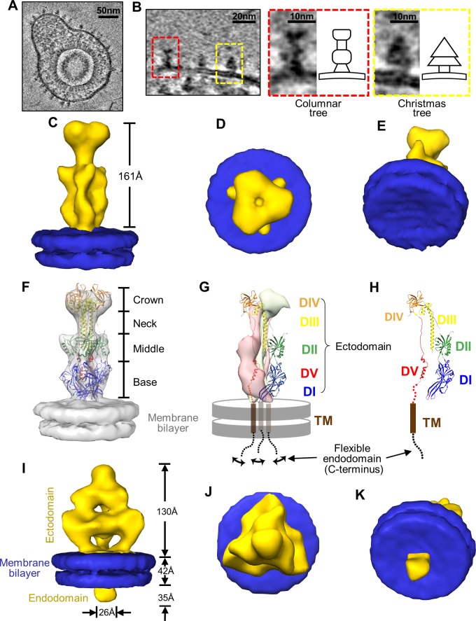
Fig 3. In situ structures of gB in the “postfusion” and prefusion conformations.
(A) A representative virion showing various glycoprotein densities on its envelope. (B) Identifications of the columnar tree-shaped (red box) and Christmas tree-shaped (yellow box) glycoprotein densities that both match the expected volume of gB (see main text). Insets are the enlargements of the two forms with their corresponding shape schematic. (C~H) Sub-tomographic average of the columnar tree-shaped glycoprotein densities, whose ectodomain matches the crystal structure of gB ectodomain in the postfusion conformation (PDB: 5CXF) [16]. The subtomographic average of the columnar-shaped densities (yellow) and segmented membrane bilayer (blue, from I) are shown either as shaded surfaces viewed from side (C), top (D) and slanted bottom (E), or as semi-transparent gray fitted with the gB ectodomain trimer crystal structure (ribbon) at the postfusion conformation (PDB: 5CXF) [16] (F). Two subunits of the gB trimer crystal structure are shown as pink and gray surfaces, while the third subunit as ribbons with its domains colored as in [16] and its transmembrane helix as brown cylinder and the C-terminal flexible endodomain as a swinging dotted lines (G). For clarity, the third subunit is shown alone in (H) with five domains (DI~DV) indicated. (I~K) Sub-tomographic average of the Christmas tree-shaped densities (yellow) and associated membrane bilayer (blue) viewed from side (I), top (J) and slanted bottom (K).

