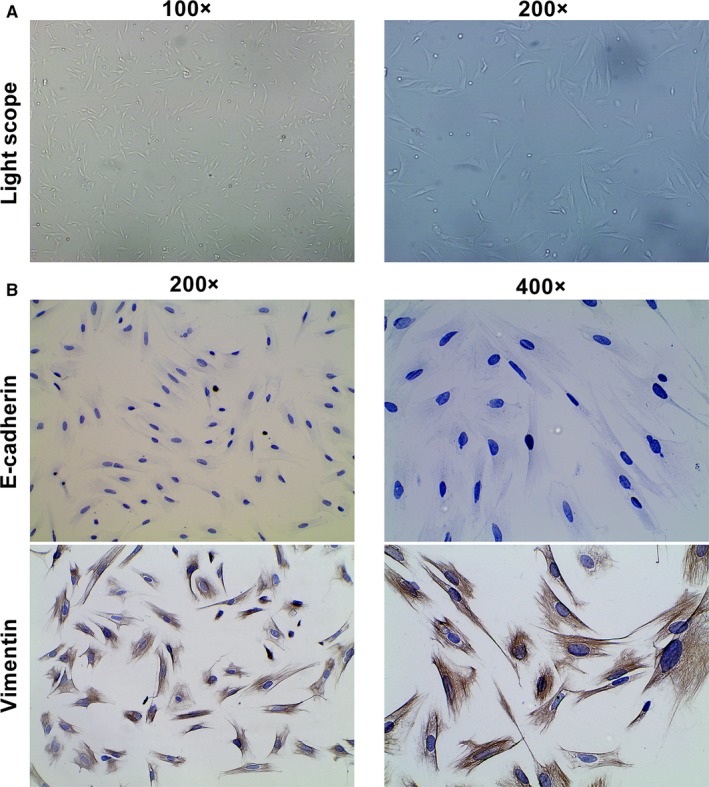
Figure 1.

Identification of the primary human endometrial stromal cells. A, Representative of morphology of cultured endometrial stromal cells. Photographs were taken at magnifications of 100× (left panels) and 200× (right panels), respectively. B, Representative of immunocytochemistry staining of E‐cadherin and vimentin protein in endometrial stromal cells. Photographs were taken at magnifications of 200× (left panels) and 400× (right panels), respectively
