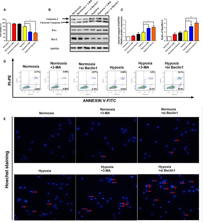
Figure 6.

Inhibition of autophagy by 3‐MA and si Beclin1 promotes apoptosis of human endometrial stromal cells under hypoxia condition. A, Representative images of the cell viability in human endometrial stromal cells after treated with 3‐MA and si Beclin1 with or without the presence of hypoxia. (B‐C) Representative Western blot of cleaved caspase‐3 and Bax/Bcl2 ratio in human endometrial stromal cells after treated with 3‐MA and si Beclin1 with or without the presence of hypoxia. D, Representative flow cytometry images of cell apoptosis in human endometrial stromal cells after treated with 3‐MA and si Beclin1 with or without the presence of hypoxia. E, Representative fluorescence images of human endometrial stromal cells stained with Hoechst 33342 fluorescent dye. Human endometrial stromal cells were treated with 3‐MA and si Beclin1 with or without the presence of hypoxia. Photographs were taken at magnifications of 200×. The protein expression levels were quantified by Image J software and normalized to GAPDH protein levels. The data are presented as the means ± SD of three independent experiments (*P < 0.05; **P < 0.01)
