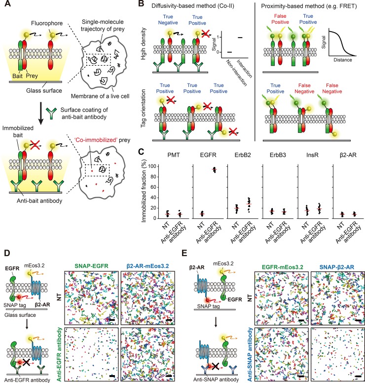
Fig 1. Membrane protein interactions are directly visualized using co-immunoimmobilization (Co-II).
(A) Schematic of the Co-II assay. The interaction between a fluorescently labeled prey protein and a bait protein is specifically probed by the co-immobilized prey produced after antibody-induced immobilization of the bait protein, which is visualized using sptPALM in single living cells. (B) Comparison between a diffusivity-based method (Co-II) and a proximity-based method (e.g., FRET). In the crowded membrane of living cells, Co-II specifically detects genuine interactions between membrane proteins, while the proximity-based methods are vulnerable to producing false positive signals because a prey and a bait are located nearby. Co-II captures membrane protein interactions independent of tag orientation, while the proximity-based methods require a careful design for donor–acceptor orientation. (C) The bait-specific immobilization using a surface-coated antibody in living cells. The immobilized fractions of PMT, EGFR, ErbB2, ErbB3, InsR, and β2-AR in multiple cells before (NT) and after anti-EGFR antibody treatment. Examined membrane proteins were expressed at a level at least 10 times higher than the expression level of EGFR to avoid the specific co-immobilization resulting from the genuine interaction with EGFR. Each dot represents single-cell data, and the red solid lines indicate the average of the immobilized fraction obtained from multiple cells (n > 10). (D–E) Illustration and trajectory maps for validation of molecule-specific immobilization in the plasma membrane of a living cell. A total of 400 trajectories are shown in each trajectory map. Scale bar, 2 μm. SNAP-EGFR was specifically and almost completely immobilized by anti-EGFR antibody treatment, whereas the immobilized fraction of β2-AR-mEos3.2 was not altered (D). Specific immobilization of β2-AR against EGFR was confirmed vice versa using SNAP-β2-AR and EGFR-mEos3.2 with anti-SNAP antibody (E). β2-AR, beta-2 adrenergic receptor; EGFR, epidermal growth factor receptor; ErbB2, erb-b2 receptor tyrosine kinase 2; ErbB3, erb-b2 receptor tyrosine kinase 3; FRET, fluorescence resonance energy transfer; InsR, insulin receptor; mEos3.2, monomeric Eos fluorescent protein variant 3.2; NT, not treated; PMT, plasma membrane targeting; SNAP, SNAP-tag; sptPALM, single-particle tracking photoactivated localization microscopy.

