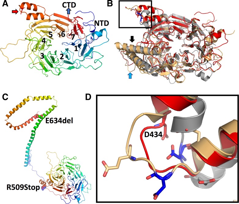
Fig 2. Modelling of human NUP88.
(A) The N-terminal domain of NUP88 reveals a seven-bladed ß-propeller with an N-terminal extension. The rainbow coloring indicates N-terminal residues in blue (NTD = N-terminal domain) and C-terminal residues of the propeller in red. Individual blades are indicated by numbers. The red arrow indicates the location of the p.D434Y point mutation (see below for details). (B) Overlay of the NUP88 model (red) with the X-ray structures of Nup82 from baker’s yeast (PDBid: 3pbp, in gray) and C. thermophilum (PDBid: 5cww; in light yellow). Significant differences between species are in blade 4 (HTH-motif; black arrow) and 5 (extended loop; blue arrow). (C) Composite model of the N- and C-terminal regions of NUP88. The presented model was generated using RaptorX with its standard settings and misses about 40 amino acid residues after the propeller region. Both, the propeller and CTD regions are colored in rainbow coloring as in (A). The individual mutations are indicated by their numbering and represented in sphere mode. (D) Magnification of the loop bearing the D434 mutation in NUP88 in stick mode. The coloring of the individual molecules is as described in (B).

