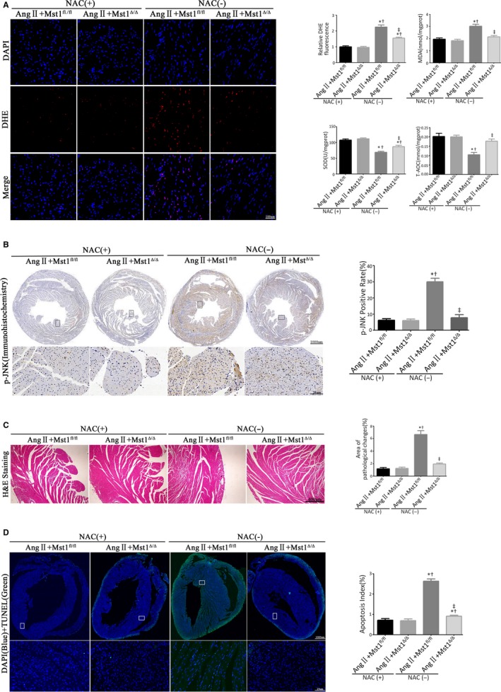
Figure 3.

Mst1 deficiency inhibits ROS‐mediated JNK phosphorylation to attenuate Ang II‐induced cardiomyocyte apoptosis in vivo. A: Measurements of ROS production (n = 6). The effects of ROS generations were measured by the DHE fluorescent probe, the levels of intracellular MDA, SOD, and T‐AOC. Histogram: Relative DHE fluorescence intensity, MDA levels (nmol/mgprot), SOD levels (U/mgprot), and T‐AOC levels (mmol/mgprot). B: Representative images of p‐JNK immunohistochemistry (n = 6). Histogram: p‐JNK positive rate (%). C: Analyse myocardial pathological changes by H&E staining (n = 6). Histogram: Areas of pathological changes (%). D: Representative images of TUNEL assay (n = 6); TUNEL (green), DAPI (blue), and cTroponin I antibody (red). Histogram: Apoptosis index. *P < 0.05 vs Ang II+Mst1fl/fl +NAC (+) group; † P < 0.05 vs Ang II+Mst1Δ/Δ + NAC (+) group; ‡ P < 0.05 vs Ang II+Mst1fl/fl +NAC (−) group
