Figure 2.

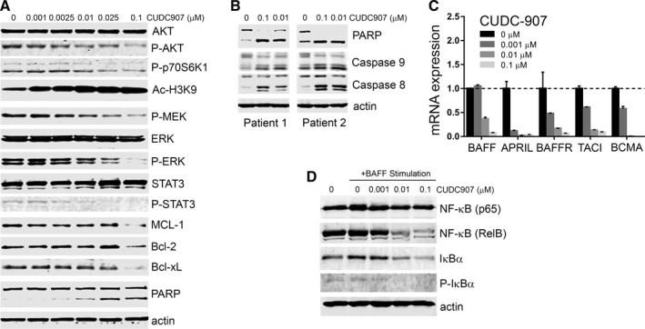
CUDC‐907 inhibits pro‐survival signals in CLL cells. A, Western blots showing protein expression in lysates of MEC‐1 cells treated with CUDC‐907 at various concentrations or DMSO as a control (0) for 12 h. AKT, p‐AKT (Ser473), p‐p70S6K (Thr389), Ac‐H3K9, ERK, p‐ERK (Thr202/Tyr204), p‐MEK (Ser217/221), STAT3, p‐STAT3 (Tyr705), MCL‐1, BCL‐2, BCL‐xL, and PARP were detected with specific antibodies. β‐actin was used as a loading control. B, Representative Western blot showing PARP, caspase‐9, and caspase‐8 cleavage in CLL primary cells from two patients. Cells were cultured as in Figure 1C and treated with different concentrations of CUDC‐907 for 12 h. C, mRNA expression levels of BAFF, APRIL, BAFFR, TCIA, and BCMA, as measured by qRT‐PCR, in CLL patient cells cultured in the presence of 10 ng/mL IL‐4 and CD154 for 24 h then treated with CUDC‐907 for 12 h. The expression of target genes was normalized to an internal control, GAPDH. Experiments were performed in triplicates and repeated at least 3 times. Data were expressed as relative to control (no inhibitor). D, Representative Western blot of lysates of CLL patient cells stimulated with BAFF (100 ng/mL) for 24 h. NF‐κB(p65), NF‐κB(RelB), IκBα, p‐IκBα, and β‐actin were detected using specific antibodies. The graph shows quantitation the Western blot bands normalized to β‐actin and expressed as relative to control (0 h) treatment
