Figure 1.

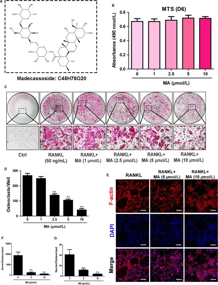
Madecassoside (MA) suppresses RANKL‐induced osteoclast differentiation in a dose‐dependent manner. (A) Chemical structure of MA. (B) The effects of the indicated concentrations of MA on BMMs were measured by an MTS assay. (C) Representative images of OCs after treatment with MA at increasing concentrations (magnification = 100×). (D) The number of TRAcP+ multinucleated cells (>3 nuclei) per well (96‐well plate) was quantitatively analysed. (E) Representative confocal images of OCs stained for F‐actin and nuclei; the images include untreated OCs and OCs treated with 5 μmol L−1 and 10 μmol L−1 Madecassoside. Scale bar = 200 μm. (F‐G) Quantification of the OCs per area and the mean number of nuclei in each cell. Data are expressed as the means ± SD; *P < 0.05, **P < 0.01, and ***P < 0.001 compared to control group
