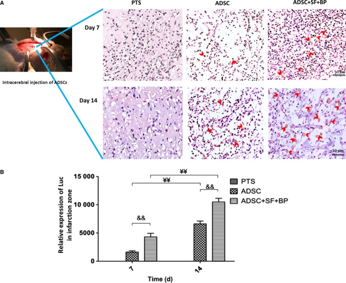
Figure 3.

Fate of ADSCs after intracerebral transplantation in the infarct zone. A, Representative images of Luc+ ADSCs detected by immunohistochemical staining at day 7 and 14. B, Quantitative result suggested a favourable effect for ADSC survival and proliferation upon SF + BP treatment. Data are expressed as means ± SD. && P < 0.01, compared with ADSC group; ¥ P < 0.05, ¥¥ P <0.01, compared with day 7. n = 6, scale bar: 50 μm
