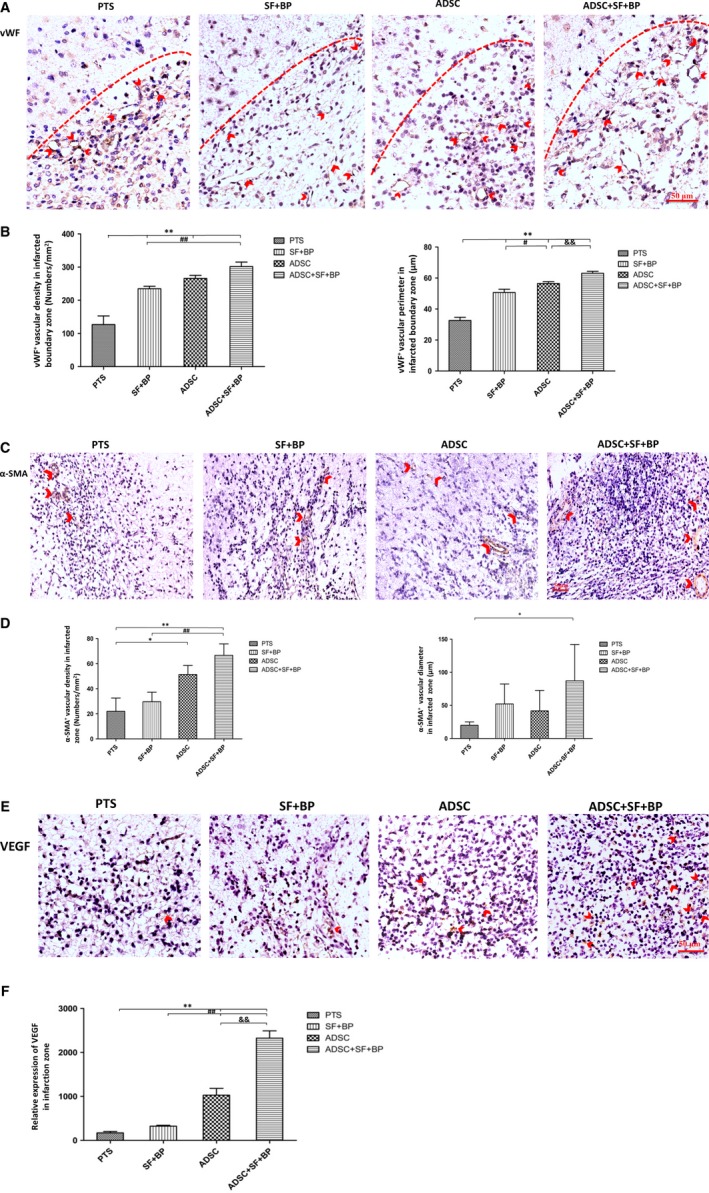
Figure 4.

Evaluations of neovascularization and VEGF expression in the infarct lesion. Representative immunohistochemistry staining images expressed brown positive signals of vWF and α‐SMA, as well as VEGF were presented (A, C, and E). Quantitative results showed that ADSC + SF + BP treatment significantly enhanced the neovascularization and VEGF expression as compared with SF + BP or ADSC treatment alone (B, D, and F). Data were expressed as means ± SD. *P < 0.05, **P < 0.01, compared with PTS group; # P < 0.05, ## P < 0.01, compared with SF + BP group; & P < 0.05, && P < 0.01, compared with ADSC group. n = 6, scale bar: 50 μm
