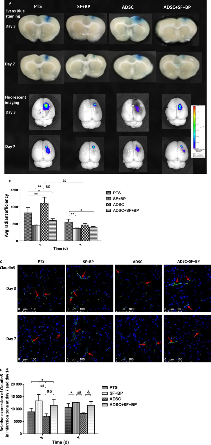
Figure 7.

Evaluational change of blood‐brain‐barrier integrity poststroke. A, Representative images of EB staining and fluorescence imaging of rat brain after stroke in different treatment groups at day 3 and 7. B, Quantitative result of EB fluorescence showed that SF + BP treatment remarkably ameliorated BBB leakage while ADSC treatment had adverse effect at day 3. However, combined treatment of ADSC + SF + BP effectively ameliorated BBB leakage at day 3‐7. (C) and (D) Representative images of claudin‐5 by immunofluorescence staining were presented and quantitative analysis showed the expression of claudin‐5 in SF + BP group was the most significant. Data were expressed as means ± SD. *P < 0.05, **P < 0.01, compared with PTS group, ## P < 0.01, compared with SF + BP group; && P < 0.01, compared with ADSC group; $$ P < 0.01, compared with day 3. N = 6. Scale bar: 100 μm
