. 2018 Dec 27;7:e41044. doi: 10.7554/eLife.41044

Figure 4. Cardiomyocyte Nrf2 is required for increase in mitochondrial antioxidant capacity with exercise.

(A) Western blots for peroxiredoxin III (PRXIII), thioredoxin reductase-2 (TRXR2) and superoxide dismutase-2 (SOD2) in heart of csNrf2KO and Flox controls after acute exercise (Ex). Mean data from n = 4–6/group are shown on the right. *p<0.05 vs respective sedentary controls (Sed); #p<0.05, ##p<0.01 vs Flox/Ex, 1-way ANOVA followed by Tukey post hoc analysis. (B) In vivo mitochondrial H2O2 levels assessed by the MitoP/MitoB ratio in hearts of csNrf2KO mice and controls after acute exercise. **p<0.01 vs Flox/Sed, 1-way ANOVA followed by Tukey post hoc analysis (n = 5–6/group). (C) MitoQ treatment improved cardiac contractile performance at peak exercise in csNrf2KO mice. n = 6–7/group. (D) and (E) The effect of MitoQ treatment on maximal running distance and time. n = 7–11/group. *p<0.05, **p<0.01 vs Flox/Ex, #p<0.05, ##p<0.01 vs csNrf2KO/Ex, 2-way ANOVA followed by Tukey post hoc analysis.
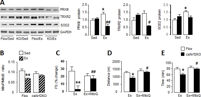
Figure 4.

Figure 4—figure supplement 1. Full images of Western blots in Figure 4.

Figure 4—figure supplement 1.

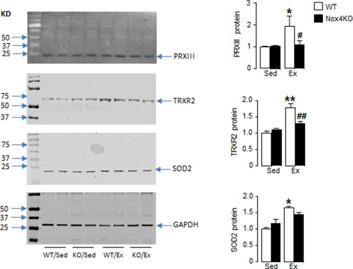
Figure 4—figure supplement 2. Nox4 is required for increase in mitochondrial antioxidant capacity with exercise.

Figure 4—figure supplement 2.

Peroxiredoxin III (PRXIII), thioredoxin reductase-2 (TRXR2) and superoxide dismutase-2 (SOD2) in the hearts of Nox4-null and littermate wild-type control mice (WT) after acute exercise (Ex). Mean data from n = 4–6/group are shown on the right. *p<0.05, **p<0.01 vs respective sedentary controls (Sed); #p<0.05, ##p<0.01 vs WT/Ex, 1-way ANOVA followed by Tukey post hoc analysis.
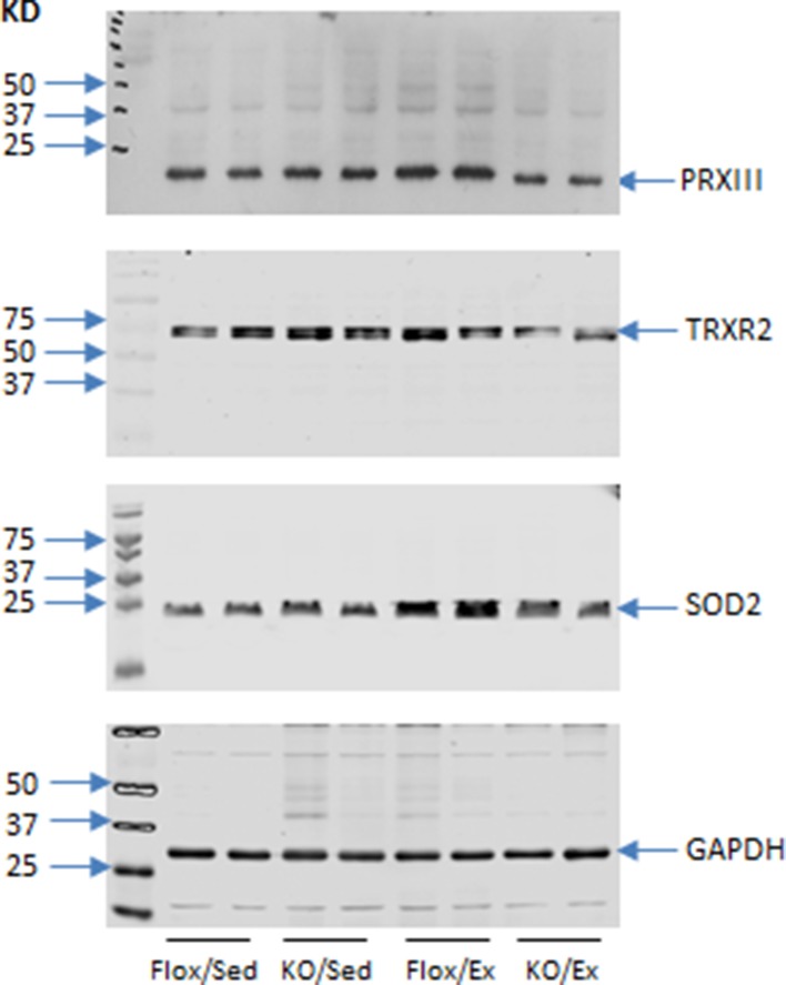
Figure 4—figure supplement 3. Mitochondrial-targeted antioxidant MitoQ improves cardiac response to exercise in csNrf2KO.

Figure 4—figure supplement 3.

Cardiac function was evaluated by conscious echocardiography prior to (sedentary, Sed) and after exercise (Ex) with or without MitoQ treatment. Mean data for heart rate (HR), left ventricular end-systolic dimension (LVESD) and left ventricular end-diastolic dimension (LVEDD) (A to C). *p<0.05, **p<0.01 vs respective sedentary controls, ##p<0.01 vs Flox/Ex, 2-way ANOVA followed by Tukey post hoc analysis (N = 6–7/group).