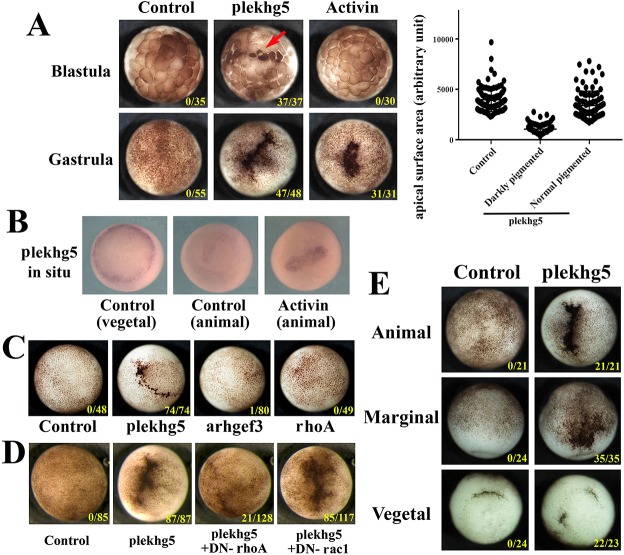
Fig. 2.
plekhg5 induces ectopic blastopore lip-like morphology in early Xenopus embryos in a Rho-dependent manner. (A) plekhg5 expression induces apical cell constriction in ectodermal cells at early blastula stages, whereas activin induces ectopic blastopore lip at the gastrula stages. The apical surface areas of cells at the blastula stages in control and the plekhg5-expressing embryos are measured and compared. The scatter plot shows a typical experiment. plekhg5 significantly reduces apical cell surfaces to about one-third that seen in control cells, with the average surface areas of 3978, 1050 and 3409 (arbitrary units) for control, apically constricted, and normal pigmented cells, respectively. Student's t-test gives a P-value of 3.5E-31 in this experiment. Red arrow indicates apically constricted cells at the blastula stages. (B) Activin induces expression of plekhg5 in the ectoderm when it induces an ectopic blastopore lip. (C) Unlike plekhg5, neither arhgef3 nor general expression of rhoA induces ectopic blastopore lip morphology in the ectoderm. (D) Dominant-negative rhoA, but not rac1, blocks ectopic blastopore lip induction by plekhg5. (E) plekhg5 induces ectopic blastopore lip morphology when injected either in the animal, the marginal zone or the vegetal regions. The doses of RNAs used are 100 pg of plekhg5, 5 pg of activin, 200 pg arhgef3, 0.5-1 ng of rhoA, DN-rhoA and DN-rac1. Numbers in each image indicate embryos exhibiting the ectopic blastopore lip-like morphology over the total number of embryos. All the experiments are repeated at least three times.

