Fig. 6.
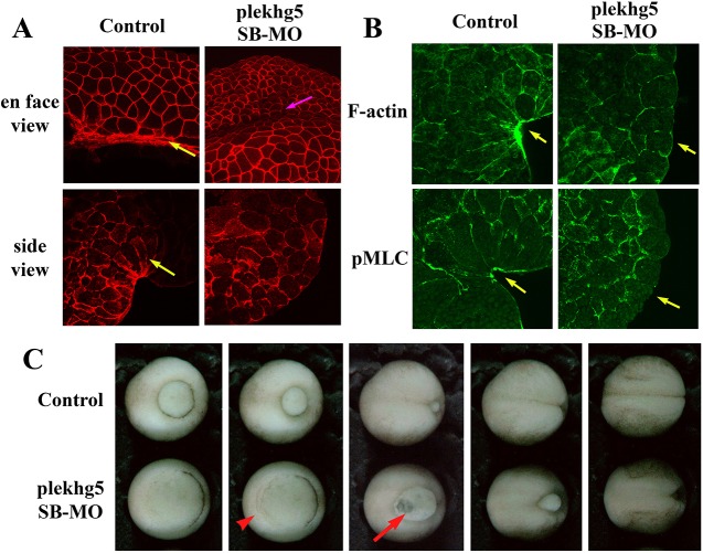
plekhg5 regulates apical actomyosin cytoskeleton in bottle cells and gastrulation movements. (A) En face and side views of control bottle cells show reduced cell surfaces and wedge-shaped morphology in gastrula embryos, respectively (yellow arrows). However, in plekhg5 morphant embryos, cells do not show great shrinkage of surface areas and only cuboidal epithelial cell shapes are seen from the side view. Despite this, internalization of surface cells appears to happen at imprecise positions in the morphant embryos, as shown by formation of a surface groove (pink arrow). (B) Both F-actin and pMLC are enriched in the apical cell cortex of the bottle cells in bisected control embryos, but no such enrichment is observed in plekhg5 morphant embryos. Yellow arrows indicate apical signals. (C) Gastrulation movements proceed in the absence of the bottle cells, as seen by accumulation of cells in the marginal region from epiboly (red arrowhead) and thinning of the vegetal mass due to rotational movements of the large endodermal cells upward and laterally (red arrow). The blastopore eventually closes in most morphant embryos, but is delayed, when control siblings reach the neurula stages. Selected still frames from a time-lapse video of gastrulating control and plekhg5 morphant embryos are shown.

