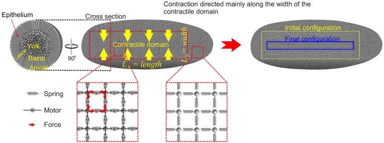
Fig. 1.
Model for gastrulation. Schematic of tissue dynamics with illustrations of the model underneath. Left: cross-section through Drosophila embryo. The embryo consists of a single layer of epithelial cells surrounding a central unstructured yolk sack. Apical surfaces face outward, basal surfaces face inward. Middle: the contractile domain (mesoderm) comprises a rectangular patch of cells some 20 cells wide and 80 cells long. Elastic elements are illustrated as springs and active forces are represented by ‘motors’ (M). Both are attached to the two adjacent ‘beads’ in parallel. Beads are assumed to feel viscous drag from the ambient environment when moving. It is assumed that forces exerted by motors are constant in time and space. Right: the contractile region shrinks anisotropically, contracting much more strongly along the width than along the length.

