Fig. 10.

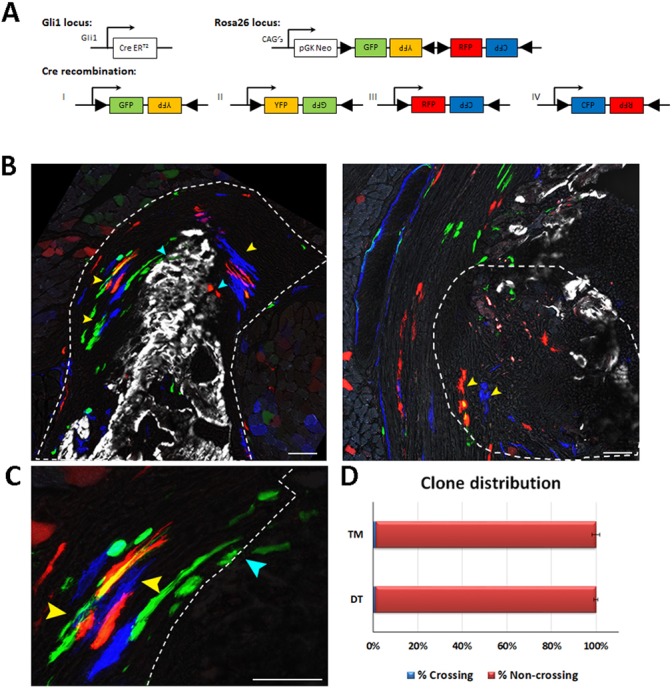
Gli1+ enthesis progenitors are compartmentalized during embryogenesis. Pulse-chase cell lineage experiments using Gli1-CreERT2;R26R-Confetti mice, in which Gli1+ progenitor cell clones were labeled. Gli1+ cells were labeled by a single tamoxifen administration at E13.5 and their descendants were followed to P14. One hour before sacrifice, mice were injected with Calcein Blue to mark mineralized tissue. (A) Schematic of possible Cre recombination outcomes. (B) Multiple clones were identified in the DT enthesis; however, the clones were restricted to either fibrocartilage or mineralized fibrocartilage and did not cross the border between the layers (yellow arrowheads). Cyan arrowheads indicate clones that crossed the border between the layers. The clones identified in the TM enthesis were restricted to the fibrous part and did not penetrate the bone. Dashed line demarcates the enthesis. (C) Magnification of DT clones in B showing crossing (cyan arrowheads) and non-crossing clones (yellow arrowheads). Dashed line marks the mineralization border. (D) Only a small percentage of identified Gli1+ clones crossed the mineralization border in the DT (1.09%±0.9) and in the TM (1.22%±1.725). Scale bars: 50 µm.
