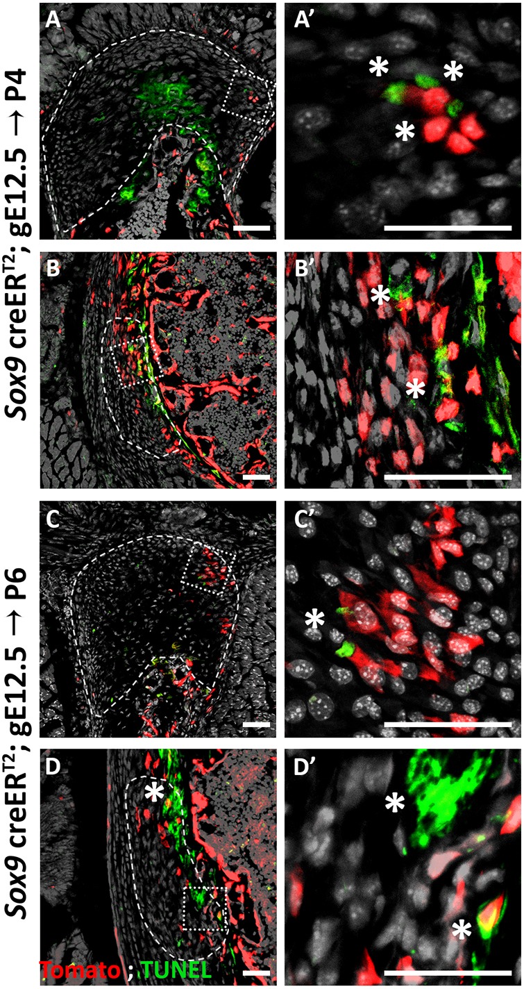
Fig. 8.

Some enthesis cells undergo cell death. (A-D′) TUNEL staining of P4 (A,B) and P6 (C,D) humeri show apoptotic cells in the DT (A,C) and in the TM (B,D). E13.5 autopod was used as a control (see Fig. S1). Dashed line demarcates the enthesis; asterisks indicate TUNEL-positive cells. A′,B′,C′,D′ are magnifications of the boxed areas in A,B,C,D, respectively. Scale bars: 50 µm.
